Ventricular Tachycardia/Ventricular Ectopics

What are ventricular tachycardias (VT) & ventricular ectopics (PVCs)?

Ventricular tachycardia (VT) is an abnormal heart rhythm that comes from the lower chambers of the heart (the ventricles).  This will often make the heart beat rapidly which can result in dizziness, palpitations and occasionally blackouts.  Ventricular tachycardia will often occur in two patient sub-groups.  The first and most common group are patients who have structural heart disease.  These patients have some structural abnormality of the heart such as a previous heart attack or a cardiomyopathy (cardiomyopathies such as hypertrophic cardiomyopathy, ARVC and familial inherited cardiomyopathies). 

Ventricular tachycardia can also occur in the setting of a structurally normal heart and this is usually a more benign problem. 

Ventricular ectopic beats are simply extra beats that arise from the lower chambers of the heart, they usually occurs as single extra beats. They are very common, and do not necessarily mean that there is any underlying heart problem.

What are the common symptoms of VT/ventricular ectopics?

VT is essentially a run of ventricular ectopics that occur in a row.  Depending on how long the episode lasts for patients can feel palpitations, dizziness and fluttering in the chest.  In severe circumstances patients can be dizzy and blackout.  Ventricular ectopics are far more common than VT.  These occur when the ventricles fire off single extra beats which patients can sense as a fluttering or a skipped/missed beat.  These are usually a benign problem and are far less serious than VT.  They are very common.

What are the causes of VT/ventricular ectopics?

Ventricular tachycardia can occur in two settings.  Firstly and most commonly are patients with structural heart disease.  Usually a scar or area of fibrosis in the heart creates areas of abnormal heart musculature and electrical conduction and this can set off the episode of VT.  In patients who have a structurally normal heart there is often nothing visible to see on an echocardiogram or a MRI scan and the heart looks completely normal.  These patients often have a “hot spot” where the VT originates from.  This is usually a less serious problem but can still require treatment.  Ventricular ectopics (PVCs) are far more common.  They can be triggered by stress, alcohol, tiredness and sometimes they can occur at random.  Ectopic burden can vary from a few extra beats a day to sometimes many thousand extra beats a day.

How do we control VT/ventricular ectopics?

Lifestyle modifications are important. Reduction in alcohol and stimulants such as caffeine/nicotine help.  If this does not control things then medication can be used.Typical medications that can be prescribed include beta-blockers or calcium channel blockers such as verapamil or diltiazem (which are only appropriate in certain types of VT).  In VT we sometimes need to use stronger drugs such as Amiodarone but we tend to reserve this drug for older patients who have significant structural heart disease.  Some patients do end up needing catheter ablation for their VT or ventricular ectopics.  This can be an extremely effective way of suppressing and getting rid of the ventricular tachycardia or the ventricular ectopic focus.  The decision to embark upon ablation depends on the severity and frequency of symptoms.  In some cases of patients who particularly suffering with VT and have significant structural heart disease we will often recommend the implantation of an ICD to protect patients from cardiac arrest.  Implantation of ICDs is not usually required in patients who have VT with a structurally normal heart who tolerate the VT well and generally these patients are treated with either drug therapy or catheter ablation only.

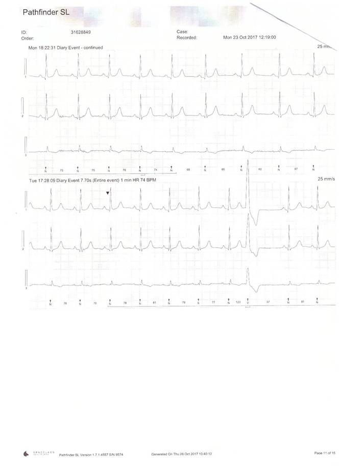
Ambulatory heart recorder trace of a patient suffering with ‘skipped beats’. Note ‘large’ beat on bottom of trace (8th beat along) is a ventricular ectopic beat or PVC

What is involved with a VT or PVC ablation?

The procedure is carried out in a cardiac catheter laboratory.  It’s usually performed under local anaesthetic and conscious sedation.  We put in between 3 and 4 catheters which are introduced via the right groin into the heart.  We generally use a complex 3D mapping system (such as CARTO or RHYTHMIA) to allow us to precisely localise the area that is causing the VT or ventricular ectopics to come from.  We then ablate that area and test to ensure we have done enough ablation to get rid of the “hot spot”.  These types of procedures can typically take 2 hours for a ventricular ectopic ablation and 3-4 hours for a VT ablation particularly if it is associated with structural heart disease.  Patients who have a structurally normal heart and have ventricular ectopics can often be discharged on the same day but patients with more advanced heart conditions such as structural heart disease who have had a more prolonged procedure are often kept in overnight.  We do occasionally do these procedures under general anaesthetic (particularly for patients who have more severe forms of VT).

Book A Consultation